 NL-BioImaging
fair-metadata-integration
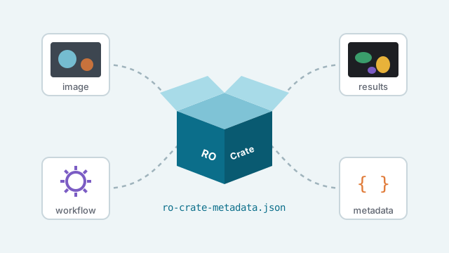
Integrate and package all metadata into a Workflow RO-Crate (NWO-funded)

Data scientist · Research Software Engineer
Dr Joost de Folter is a multidisciplinary data scientist and Research Software Engineer specialising in microscopy data science, machine learning, image analysis, modelling and visualisation. He divides his time between the Francis Crick Institute in London, where he develops image analysis and registration software for large-scale microscopy research, and NL-BioImaging in the Netherlands, where he works on FAIR bioimaging data infrastructure, OME-Zarr conversion and metadata standards across the national network. He has extensive experience developing automated and reproducible solutions across biomedical research, healthcare and AI, with a particular interest in open standards, open science and translating complex scientific challenges into practical computational solutions.
NL-BioImagingIntegrate and package all metadata into a Workflow RO-Crate (NWO-funded)
 NL-BioImaging
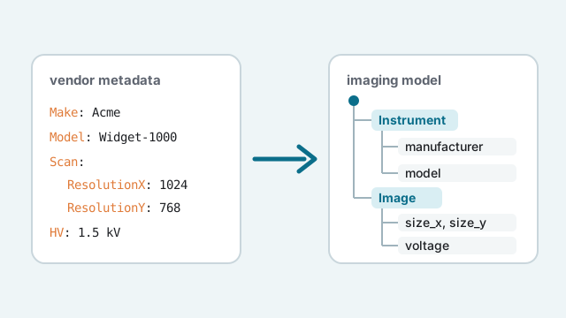
Convert acquisition metadata to custom model (NWO-funded)
 NL-BioImaging
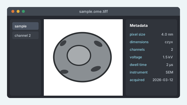
napari tiff reader with extended metadata (NWO-funded)
 NL-BioImaging
BIOMERO.analyzer - A python library for easy connecting between OMERO (jobs) and a Slurm cluster (NWO-funded)
 NL-BioImaging
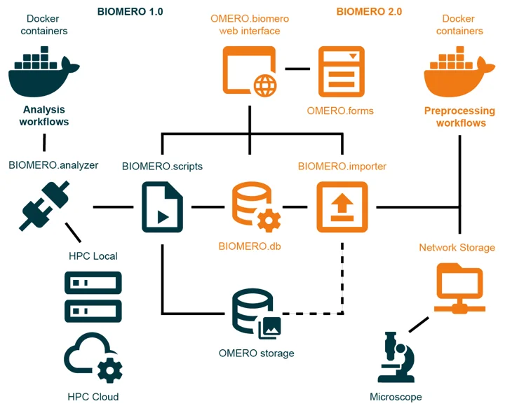
NL‑BIOMERO delivers a full containerized stack to run OMERO together with the BIOMERO 2.0 framework. It provides Docker/Podman configurations and Compose files to deploy OMERO + BIOMERO subsystems (importer, analyzer, OMERO.web plugin, databases, and auxiliary services) — the recommended starting point for a FAIR‑oriented bioimaging setup. (NWO-funded)
 folterj
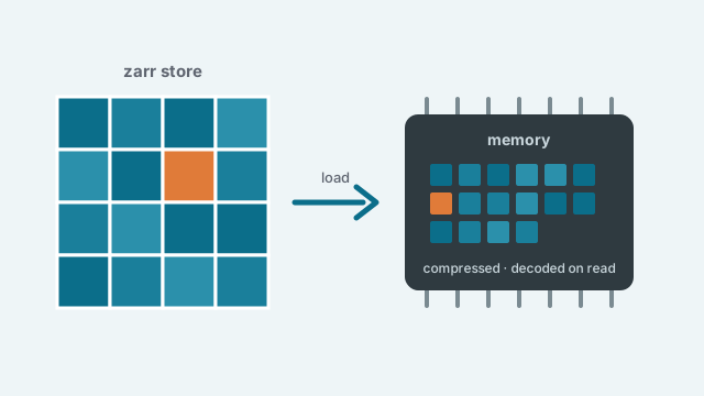
Zarr in-memory compressed data loader
 NL-BioImaging
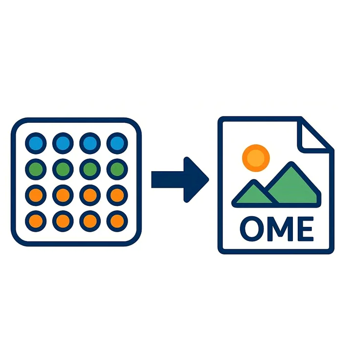
Convert db image format to Ome-Tiff and Ome-Zarr (NWO-funded)
 FrancisCrickInstitute
Ome(ro) SLide Image Conversion and Compression pipline (OmeSliCC)
 ccp-volume-em
Community best practice guide (UKRI-funded)
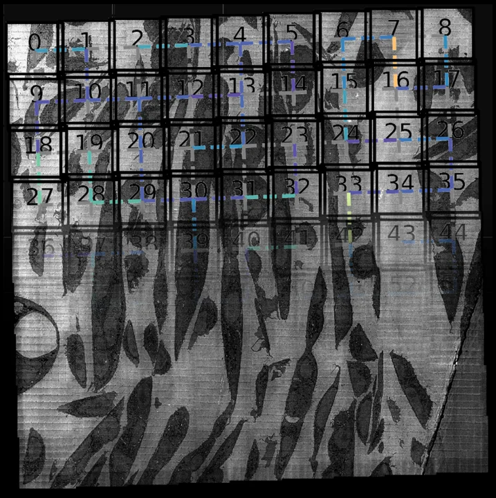 ccp-volume-em
Image registration using multiview-stitcher (UKRI-funded)
 NL-BioImaging
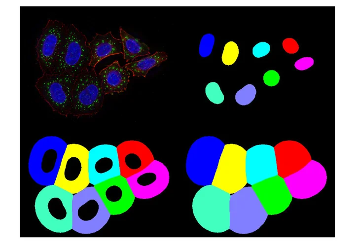
FAIR organelle segmentation pipeline (NWO-funded)
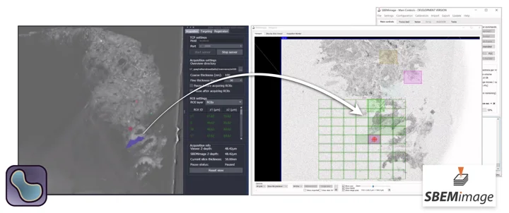 FrancisCrickInstitute
Multimodal SBEM acquisition targeting (MRC-funded)
 FrancisCrickInstitute
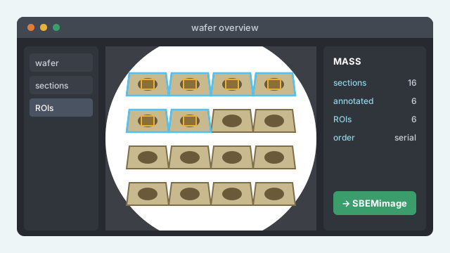
Microscopy Array Section Setup napari plugin (CZI-funded)
 SBEMimage
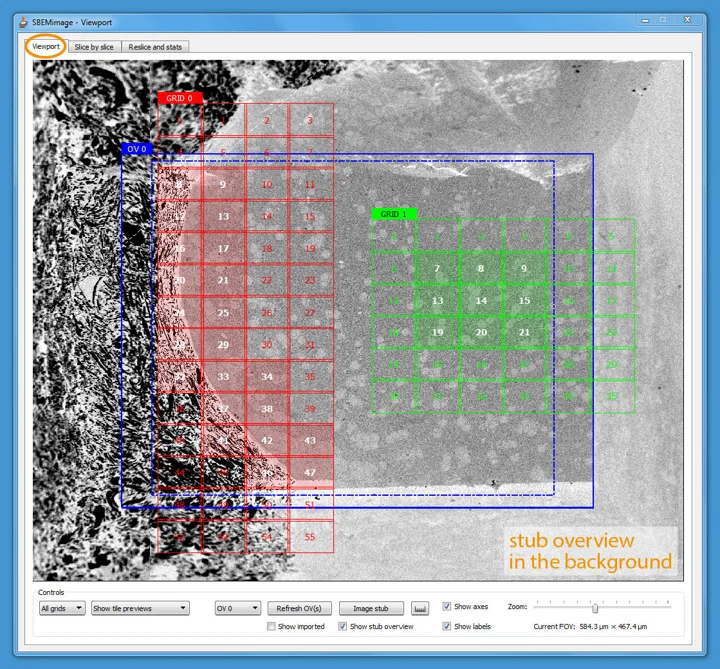
Versatile acquisition software for serial block-face electron microscopy
 folterj
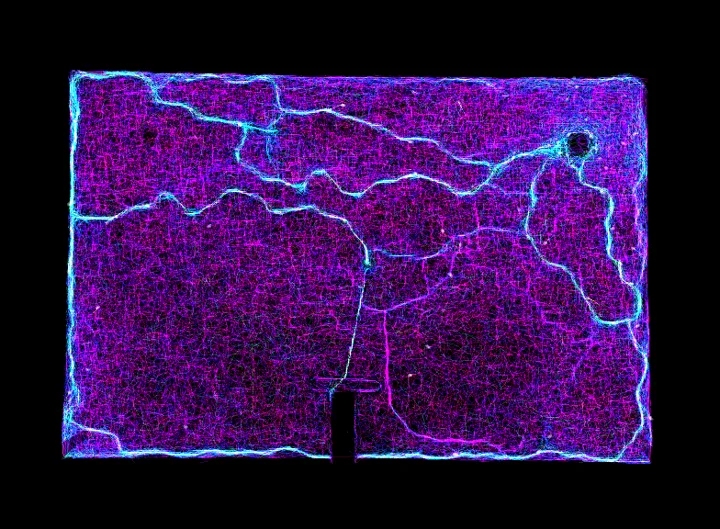
Image processing tool focusing on biological applications
 FrancisCrickInstitute
Deep learning segmentation of the nuclear envelope in electron microscopy, trained on citizen science annotations
 folterj
Analyses and validates binary IEEE 11073 messages in explorable formats
 folterj
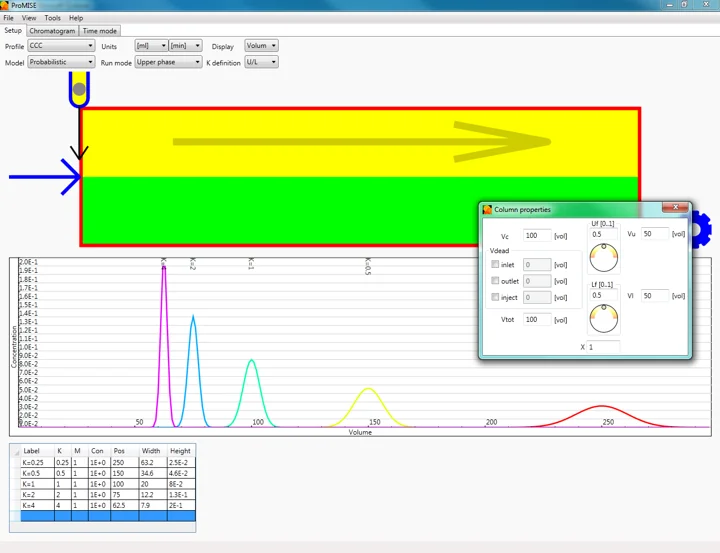
Probabilistic Model for Immiscible Separations and Extractions 2
 fideus-labs
 ome


